Research
We are interest on every medical image modality analysis, with a string knowledge and know-about with MRI. Part of our research lines of interest are:
 Denoising
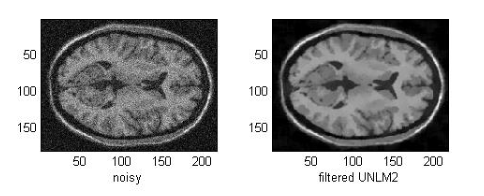
Magnetic Resonance (MR) images are affected by random noise which limits the accuracy of any quantitative measurements from the data. Therefore, filters for random noise removal in MR magnitude images are studied, as the parametric filter Non-Local Means (NLM).
More information here.
Inhomogeneity Correction
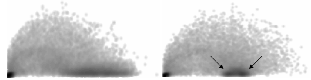
MR images are commonly affected by signal inhomogeneities that hampers the obtention of quantitative information from them. In the last years, the correction of this inhomogeneity (also referred as bias correction) has gain a great attention and methods has been developed inside of the group.
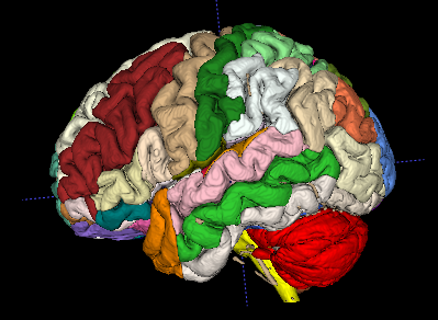
Brain Segmentation
Medical image segmentation has evolved significantly over the years. The research group has developed pipelines like the volbrain platform, which offer fully automatic volumetric brain analysis.
However, recent advancements in deep artificial intelligence techniques have led to the development of pipelines like DeepHIPS, DeepICE, and AssemblyNET, which are powerful ensembles of convolutional neural networks for 3D Whole Brain MRI segmentation. The group has also focused on pathologies and lesions like Alzheimer’s, Parkinson’s, and multiple sclerosis, with tools like pBrain, HAVAs, DeepLesionBrain, and AssemblyNet-AD, developed to address these conditions and facilitate accurate lesion segmentation.
Superrresolution
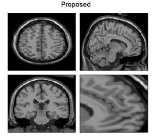
The resolution of MRI images is limited by several factors, such as hardware or time constraints. In many cases, acquired images have to be oversampled to fit a specific resolution; in these cases, image interpolation techniques have traditionally been applied. However, traditional interpolation techniques are not capable of recovering the high frequency information from the underlying high resolution data. The group has worked on the problem with developments such as Non-Local MRI Upsampling and currently applying Deep Learning to the problem.
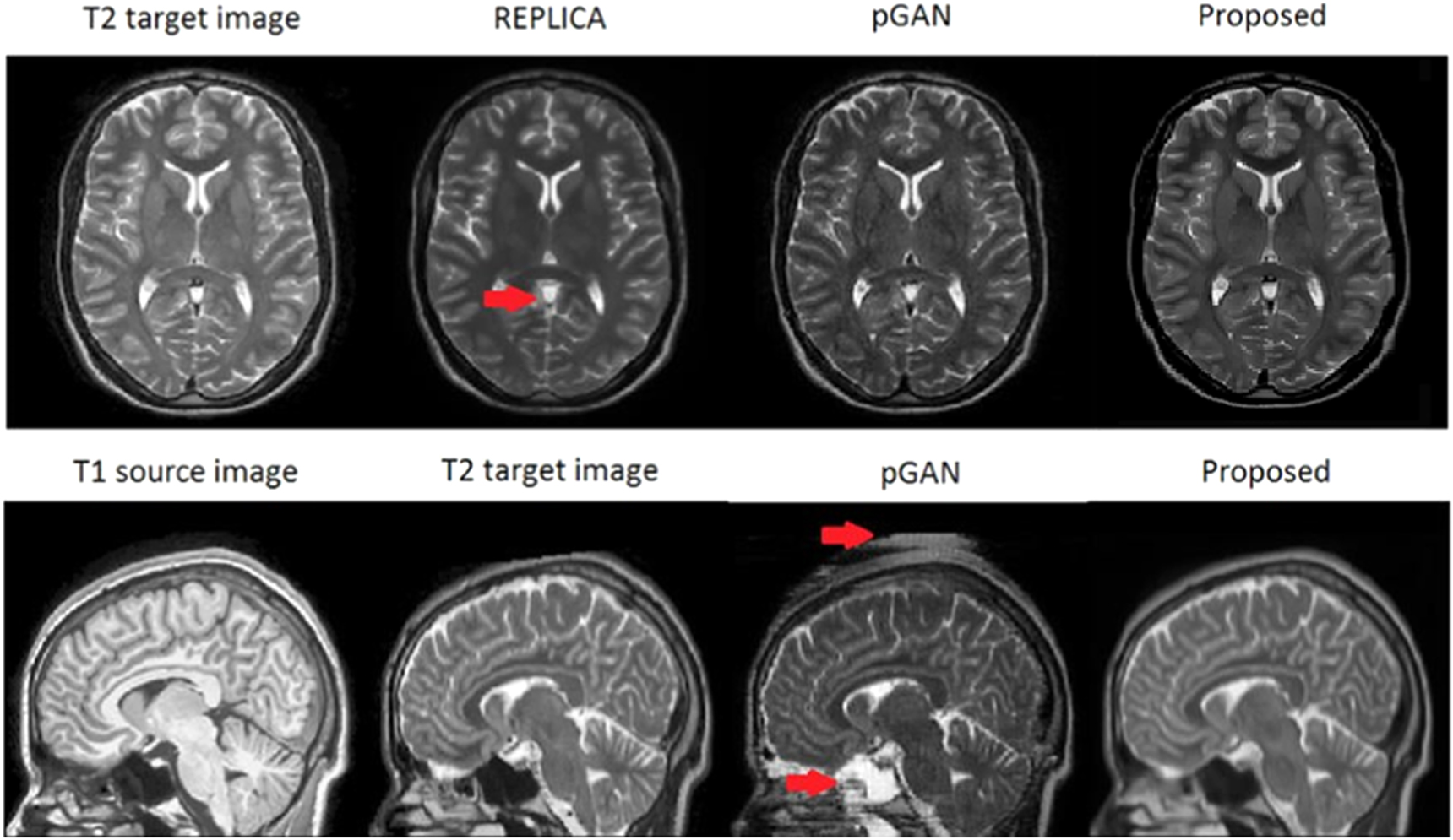
Synthesis
MRI analysis tasks can benefit from generating multiple image types or contrasts, which can enhance segmentation and registration. However, acquiring multiple contrasts can be challenging due to time constraints and patient comfort. Contrast synthesis offers a promising approach to generate additional image types. In our recent research we have developed tools like Deep Learning-based MRI contrast synthesis using full volume prediction, which use advanced deep learning techniques to synthesize alternative image types, expanding available information and improving the overall MRI workflow.
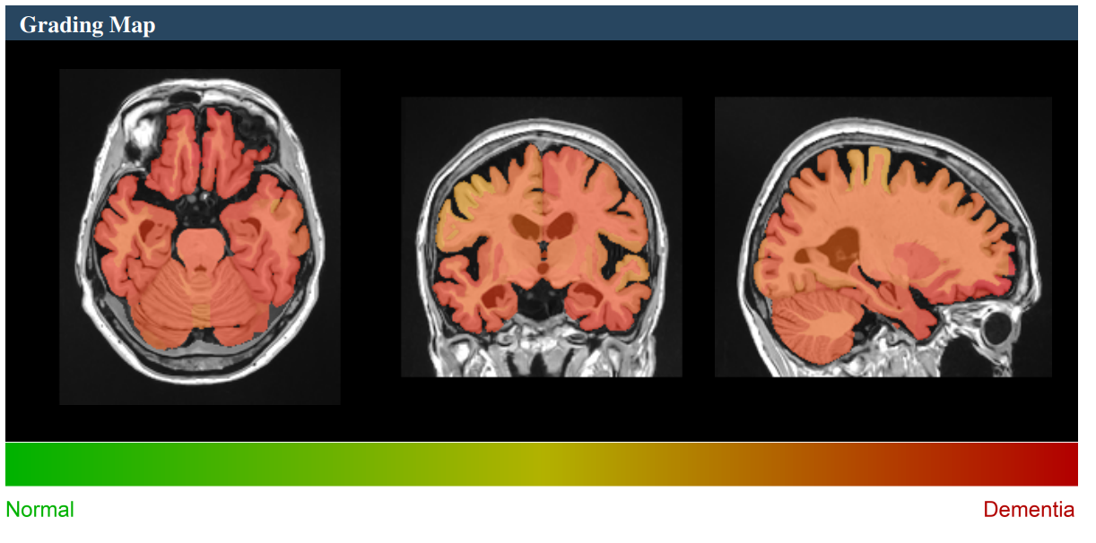
Diagnostics
Over the past ten years, computer-aided diagnosis has seen substantial growth as a research area. This field focuses on creating novel methods for analyzing images to support medical professionals in interpreting diagnostic images. Typically, it involves an automated process that includes preprocessing, extracting specific features, and classifying these features. Moreover, computers have become indispensable in tasks like comparing numerous images simultaneously or detecting subtle anatomical changes induced by illnesses.
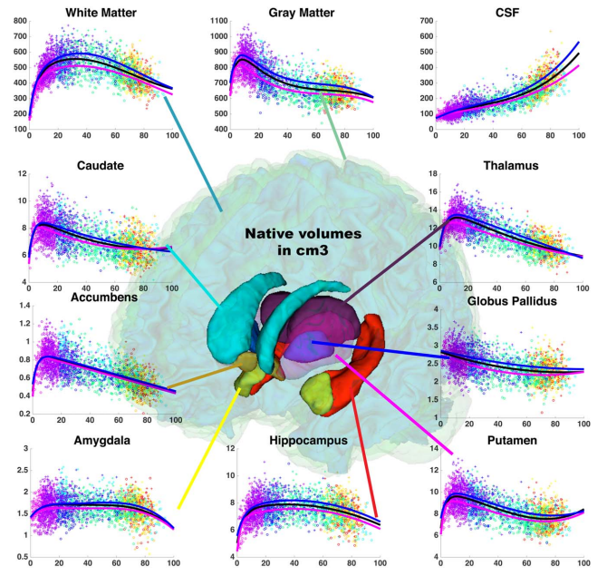
Lifespan
To answer questions related to the evolution of diseases and to be able to detect biomarkers, it is necessary to have models of ageing and to be able to study disease trajectories. To this end, we proposed an innovative way by inferring brain structure model across the entire lifespan using a massive number of MRI (N=4329) Towards a unified analysis of brain maturation and aging across the entire lifespan: A MRI analysis